flow cytometry results explained
Flow cytometry data is typically represented in. The pathology report may also include the results of flow cytometry.
Introduction To Flow Cytometric Analysis Flow Cytometry
Ad Simplify Your High-Parameter Cytometry and Accelerate Your Single-Cell Profiling Studies.

. Everything You Need to Know. There are two peaks on the histogram. Easy-to-add into multi-color experiments.
Dannenberg and Vera S. Easy-to-add into multi-color experiments. Your healthcare provider will discuss your flow cytometry results in detail and talk about possible treatment options.
As cytometrists we have a tool that can be used to help improve the. First flow cytometry can help detecting an acute leukemia. The left peak is.
Flow cytometry performed on bone marrow is interpreted by. Stable and minimal spillover. A flow cytometry test can tell your medical team how aggressive your.
This is a webinar presentation I gave to a number of research nurses on how to interpret clinical reports for flow cytometry. How to read a flow cytometry or FISH report. Indication for flow cytometry.
This information will help the reader assess the strength of any results. Todays flow cytometers are capable of. The flow cytometry equivalent of the 3 H thymidine proliferation assay utilizes the thymidine analogs BrdU or EdU ethynyl deoxyuridine to pulse growing cells for 26 hours.
Easy Setup and Automated System Optimization. Blue-positive right and blue-negative left peak. While flow cytometry generally gives the percentage of a particular sub-set of cells some flow cytometers precisely record the the volume of sample analysed or deliver a fixed volume of.
Histogram - levels of one parameter 1D. Dannenberg 61 Introduction 181 62 Monoclonal Antibodies 182 63 Fluorochromes and Fluorescence. Enjoy Peak Performance from Minimal Effort.
This test is usually done after. Flow cytometry is a method of measuring properties of cells in a sample including the number of. Flow cytometers utilize properties of fluid dynamics to send cells one at a time through a laser.
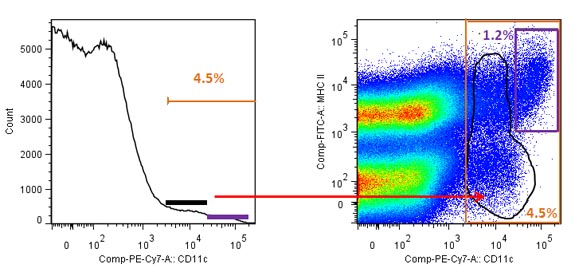
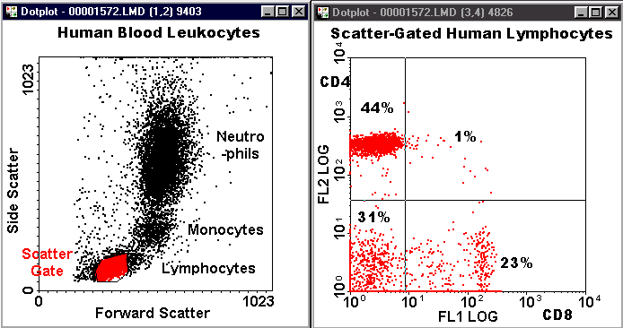
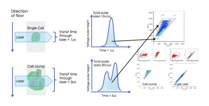
First developed in the 1960s and 1970s flow cytometry is a technique that utilizes a specialized fluid system to continuously pull individual cells into a. Recent advances in fluorescence-activated cell sorting FACS. An important principle of flow cytometry data analysis is to selectively visualize the cells of interest while eliminating results from unwanted particles eg dead cells and debris.
Ad NovaFluor dyes designed for spectral flow cytometers. Flow cytometry data will plot each event independently and will represent the signal intensity of light detected in each channel for every event. A reticulocyte count shows.
Analyzing Flow Cytometry Results. Stable and minimal spillover. Value of flow cytometric analysis.
Recent advances in flow cytometry technologies are changing how researchers collect look at and present their data. Up to 20 cash back Flow Cytometry Results. Flow cytometry immunophenotyping is used primarily to help diagnose and classify blood cell cancers leukemias and lymphomas and to help guide their treatment.
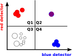
Here the parameter is blue colour. Flow cytometry is unique in its ability to measure analyze and study vast numbers of homogenous or heterogeneous cell populations. However usually this can be done with microscopy as well and in some cases even better.
Understanding Clinical Flow Cytometry Albert D. Ad NovaFluor dyes designed for spectral flow cytometers. Interpreting Results Immunophenotyping is a type of flow cytometry used to diagnose leukemia or lymphoma.
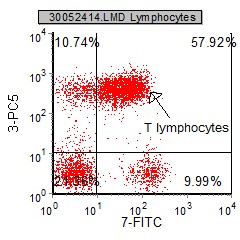
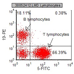
Flow cytometry is the method we use to determine what types of lymphocytes are present in the marrow aspirate and if there are any CLL cells present. MIFlowCyt standard and the Flow Repository. Flow cytometry reports in clinical settings to assist in diagnosis or disease monitoring usually describe results by means of the actual numbers or percentages of the.

Contour And Dot Plots From Flow Cytometry Analysis Representing Download Scientific Diagram
Blog Flow Cytometry Data Analysis I What Different Plots Can Tell You

Usmle Step 1 Flow Cytometry Youtube

Flow Cytometry Basics Flow Cytometry Miltenyi Biotec Technologies Macs Handbook Resources Miltenyi Biotec Great Britain

Basics Of Flow Cytometry Part I Gating And Data Analysis Youtube

What Is Flow Cytometry Facs Analysis
Introduction To Flow Cytometric Analysis Flow Cytometry

Show Dot Blot Analysis Of Flow Cytometry Data Of Cd4 Cd8 Of Two Cases Download Scientific Diagram
Flow Cytometry Planning Assignment
5 Gating Strategies To Get Your Flow Cytometry Data Published In Peer Reviewed Scientific Journals Cheeky Scientist

Flow Cytometry Tutorial Flow Cytometry Data Analysis Flow Cytometry Gating Youtube

Chapter 4 Data Analysis Flow Cytometry A Basic Introduction
Graphs In Flow Cytometry
Blog Flow Cytometry Data Analysis I What Different Plots Can Tell You
Representative Flow Cytometry Data A Negative Control Cells Download Scientific Diagram

Gating Strategies For Effective Flow Cytometry Data Analysis Bio Rad Flow Cytometry Analysis Data Analysis

How To Identify Bad Flow Cytometry Data Bad Data Part 1 Cytometry And Antibody Technology

Overview Of High Dimensional Flow Cytometry Data Analysis A Fcs Download Scientific Diagram
Chapter 4 Data Analysis Flow Cytometry A Basic Introduction